What is a Neck MRI?
Among magnetic resonance imaging methods, a neck MRI provides detailed information about the vertebrae, muscle structures, nerves, blood vessels, and soft tissues in the neck region. It is a safe method since it does not use radiation and offers high-resolution images. This allows for easy examination of conditions such as herniated discs, tumors, vascular blockages, or nerve compression. We listen to our patients’ complaints and provide imaging with advanced MRI devices to reach an accurate diagnosis.
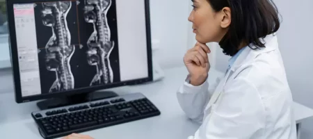
How is a Neck MRI Performed?
The patient lies on their back in the MRI device, and the neck region is imaged with special coils. The patient must remain completely still during the procedure; otherwise, the images may become blurred. Hearing loud noises during the scan is normal, and headphones or earplugs are used to ensure patient comfort. If necessary, a contrast agent can also be applied. We place great importance on patient comfort during neck MRI scans and complete the procedure as safely and quickly as possible.
When is a Neck MRI Recommended?
An MRI is preferred in cases of severe neck pain, numbness radiating to the shoulders, dizziness, muscle weakness, or suspicion of vascular blockage. It is also very useful for examining herniated discs, masses, tumors, infections, and abnormalities in vascular structures. Especially after trauma or before surgery, doctors obtain detailed information with a neck MRI. We perform these imaging studies quickly and ensure that the results are reported accurately.
Is a Neck MRI Harmful?
Magnetic resonance methods do not use X-rays or radiation, so they are not harmful. Therefore, repeated scans do not pose a significant risk. However, there may be restrictions for patients with pacemakers, metal implants, or prostheses. MRI is also not recommended during the first trimester of pregnancy. We review each patient’s medical history to ensure the procedure is performed safely and appropriately.
How Long Does a Neck MRI Take?
The duration of the scan depends on the patient’s condition, the level of imaging detail required, and whether a contrast agent is used. Generally, it takes 20 to 40 minutes. Some specialized examinations may take slightly longer. We use modern devices to shorten scan times as much as possible while obtaining clear and reliable results, providing patients with both comfort and efficiency.
How Soon Are Neck MRI Results Available?
The results of a completed neck MRI are usually delivered to the patient the same day or within a few days. The timing depends on the level of detail in the images, the preparation of the report, and the reviewing doctor. In urgent cases, results are prepared and shared much faster. We evaluate the obtained images with our radiology specialists to minimize patient waiting times, allowing the treatment process to start promptly and improving quality of life.
What Can Be Seen on a Neck MRI?
MRI images provide detailed examination of the neck vertebrae, spinal cord, muscle structures, nerves, blood vessels, and soft tissues. This examination allows for easy detection of herniated discs, tumors, vascular narrowing, nerve compression, edema, inflammation, or masses. It also provides detailed information about the structure of the neck vessels and blood flow. We use the most advanced MRI devices to make every detail visible, offering reliable data for accurate diagnosis.
Precautions Before a Neck MRI
Patients should observe some precautions before the scan. No metal objects should be on the body; necklaces, earrings, watches, or belts must be removed. If a contrast agent will be used, patients may be asked to come on an empty stomach. Those with kidney problems or allergies should share this information prior to the procedure. We provide detailed guidance to our patients before the scan to ensure it is safe and comfortable.
Which Conditions Can Be Detected with a Neck MRI?
MRI examination allows early detection of many neck conditions. Herniated discs, tumors, vascular blockages, nerve compressions, cysts, inflammations, and spinal cord injuries can be clearly visualized. Narrowing of neck vessels, aneurysms, or circulation disorders can also be identified. We perform these examinations to help patients reach the correct diagnosis and support the treatment process reliably.
Neck MRI Prices 2026
Costs may vary depending on the technology of the device used, the detail of the scan, and whether a contrast agent is applied. There is a price difference between a standard single-session neck MRI and a contrast-enhanced study. If you are having the scan at a private institution, device strength, contrast, and reporting time can affect the price. An average range of 3,000–12,000 TL is reasonable.
Contact us for pricing, as every patient’s needs are different and the examination is planned individually. Our goal is to provide high-quality imaging services at reasonable conditions.
Is Neck MRI Performed with Contrast?
In some cases, a contrast agent is used, while in others it is not. The contrast agent is administered intravenously and helps to visualize blood vessels, tumors, or inflammatory areas more clearly. However, not every neck MRI requires contrast. The doctor decides based on symptoms and examination needs. We inform patients before the procedure and safely perform contrast-enhanced imaging when necessary.
Why is Neck MRI Performed?
This method provides detailed information about muscles, vessels, nerves, vertebrae, and the spinal cord in the neck. It is preferred when there is severe neck pain, numbness, dizziness, muscle weakness, or suspicion of vascular blockage. It is also used for tumor investigations, suspected herniated discs, infections, or pre-surgical preparation. We provide the clearest images to reach an accurate diagnosis and obtain data that guide treatment.
Why Do Doctors Request a Neck MRI?
Doctors request a neck MRI to obtain more detailed information based on the patient’s complaints. This imaging method is particularly preferred for herniated discs, nerve compressions, vascular narrowing, tumors, or certain neurological conditions linked to the brain. Early diagnosis facilitates treatment for many conditions. We provide reliable and fast services with modern devices so patients can reach a definitive result as soon as possible.
For What Purpose is Neck MRI Done?
A neck MRI provides detailed imaging of vertebrae, nerves, blood vessels, muscles, and soft tissues. The purpose is to determine the underlying cause of a patient’s complaints and reach an accurate diagnosis. It is used in cases of dizziness, neck pain, numbness radiating to the arms, muscle weakness, or suspicion of vascular blockage. It is also an important tool for tumor investigations, assessing infections, and pre-surgical planning. We perform these scans reliably to support the treatment process.
Which MRI is Done for a Herniated Neck Disc?
For suspected cervical disc herniation, an MRI focused on the spine and spinal cord is preferred. This examination clearly shows the location, size, and pressure on nerve roots caused by the hernia, helping to identify the source of pain and numbness. We perform MRIs for neck herniation with advanced devices, providing clear images to support accurate diagnosis.
Why is Contrast-Enhanced Neck MRI Done?
Contrast-enhanced neck MRI may be necessary in some cases. It is particularly used for examining blood vessels, determining tumor boundaries, and detecting inflammatory areas. The contrast agent helps produce clearer and more detailed images. Not every patient requires contrast MRI; the doctor decides based on symptoms. We provide safe and comfortable contrast-enhanced MRI procedures.
Does a Neck Hernia Show on MRI?
MRI is one of the most effective imaging methods for detecting neck herniation. It clearly shows the level of the hernia, whether it is pressing on nerves, and its effects on the spinal cord. This helps determine whether surgery is necessary or which treatment method should be applied. We provide detailed imaging to detect neck herniation early and guide the treatment process safely.
What Does a Neck MRI Show?
A neck MRI examines the spine structure, spinal canal, blood vessels, muscles, and nerves in detail. This allows clear visualization of hernias, nerve compressions, cysts, tumors, vascular blockages, edema, or inflammation. High-resolution images reveal even small structural changes. We obtain the most accurate results from neck MRI images and guide patients’ treatment reliably.
Why is a Neck MRI Requested?
A doctor may request a neck MRI to obtain detailed information based on a patient’s complaints. Persistent neck pain, numbness radiating to the shoulders and arms, dizziness, muscle weakness, or nerve compression are reasons for this examination. It is also preferred for tumor investigation, post-trauma evaluation, and pre-surgical planning. We perform neck MRI scans quickly and reliably, helping reach a diagnosis as soon as possible.
What Are the Symptoms of Neck Cancer?
Neck cancers can show different symptoms. Common signs include persistent pain, a palpable mass in the neck, difficulty swallowing, hoarseness, pain radiating to the ears, and swollen lymph nodes. Weight loss, fatigue, and chronic cough may also accompany in some cases. Early diagnosis is crucial, and such symptoms should not be ignored. We use advanced imaging methods like neck MRI to help patients reach an early diagnosis.
Should Clothing Be Removed for Neck MRI?
During the scan, patients should not wear clothing or accessories containing metal. Zippers, buttons, bras with wires, necklaces, or watches are removed as they can be affected by the magnetic field. Patients are usually provided with a hospital gown for the procedure. We carefully prepare patients beforehand, prioritizing their comfort.
Where Does Nerve Compression in the Neck Radiate?
Nerve compressions in the neck can cause pain and numbness radiating to the arms, shoulders, and sometimes fingertips. Muscle weakness, difficulty gripping, and decreased reflexes may also occur. Symptoms vary between individuals but generally start in the neck and radiate toward the arm. We perform detailed MRI examinations to detect nerve compressions and guide accurate treatment.
How is a Neck MRI Performed?
Patients lie on their backs, and special coils are placed on the neck. It is crucial to remain still during the scan to avoid image distortion. Loud noises are heard during the procedure, so headphones or earplugs are used for comfort. If necessary, contrast agent is applied for more detailed imaging. We perform neck MRI scans with modern devices, providing a safe and comfortable experience for patients.
Which Doctor Should Be Visited for a Neck MRI?
Patients experiencing neck pain, numbness, dizziness, or muscle weakness usually consult a neurosurgeon or neurologist. For structural problems, herniated discs, or muscle-related issues, an orthopedist may also evaluate the patient. If the doctor needs more detailed information after examination and history, a neck MRI may be requested. We perform these imaging studies quickly and reliably, ensuring results reach the relevant specialists promptly.
Does Nerve Compression in the Neck Show on MRI?
Magnetic resonance imaging clearly shows the areas through which nerves pass and the spinal canal, allowing nerve compressions to be identified. MRI images reveal pressure on nerve roots, edema, or narrowing caused by hernias. This helps determine the location and severity of nerve compression. We prepare detailed MRI examinations for patients with suspected nerve compression and support accurate treatment planning.
Does Stress Affect the Neck?
Intense stress causes muscle tension, most notably in the neck. Continuous tension leads to muscle stiffness, headaches, and pain radiating to the shoulders, which are typical examples of stress affecting the neck. Prolonged stress can make neck pain chronic. Therefore, regular exercise, correct posture, and stress management are important. We provide patients with not only imaging but also recommendations to improve their quality of life.
What Are the Symptoms of Neck Osteoarthritis?
Neck osteoarthritis occurs with aging or excessive load on the cartilage between vertebrae. The most prominent symptom is pain and restricted neck movement. Pain may also radiate to the shoulders and arms, along with numbness, muscle weakness, and dizziness. Advanced stages may lead to nerve compression. We clarify the condition with MRI imaging for patients suspected of having neck osteoarthritis and support access to the correct treatment.


