What is Multiparametric MRI?
Multiparametric MRI is one of the advanced magnetic resonance imaging methods obtained by using different MRI techniques simultaneously. While standard MRI only provides information about anatomical structure, multiparametric MRI can also examine tissue structure, function, and biological properties. In addition to anatomical images, additional sequences such as diffusion, perfusion, and spectroscopy provide cellular-level information. It has become one of the most commonly used methods, especially in the diagnosis of prostate cancer. It also allows detailed evaluation of the brain, liver, and other organs. Its ability to detect suspicious areas within the tissue earlier and more clearly than other methods makes it one of the important diagnostic tools of modern medicine.
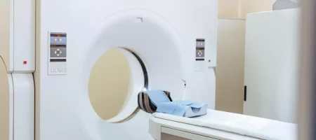
How is Multiparametric MRI Performed?
The scan is performed with standard MRI machines but with special protocols. The patient lies in a supine position in the MRI machine and must remain completely still during the procedure. Multiparametric MRI is performed by applying different sequences consecutively. First, high-resolution anatomical images are obtained, then cellular structure with diffusion MRI, blood supply with perfusion MRI, and in some centers biochemical contents with spectroscopy are evaluated. The scan takes longer than standard MRI and usually lasts between 30 and 60 minutes. The use of contrast material may vary depending on the details of the examination. The procedure is painless and does not involve radiation.
For Which Diseases is Multiparametric MRI Requested?
Its most common use is in prostate cancer. Multiparametric MRI allows detailed examination of the prostate tissue, identification of suspicious areas, and guidance for biopsy. It is also used in brain tumors, liver lesions, and some kidney diseases. In the brain, the relationship of tumor tissue with its surroundings, its blood supply, and metabolic structure can be better understood with this method. In the liver, multiparametric MRI can be preferred to distinguish between benign and malignant lesions. In general, it is one of the most reliable methods for distinguishing suspicious masses, grading tumors, and planning treatment.
Why Multiparametric MRI?
Because classical imaging methods do not always provide sufficient information. Especially in diseases such as cancer that need to be diagnosed early, multiparametric MRI reveals not only the shape and size of tissues but also their biological properties. Thus, more definitive findings are obtained as to whether the lesions are benign or malignant. It also helps to prevent unnecessary biopsies and allows targeted biopsy. It is also of great importance in treatment planning and evaluating the response to treatment.
Who Should Have Multiparametric MRI?
This test is particularly applied to men at risk of prostate cancer. Multiparametric MRI is performed in patients with high PSA levels, family history, or suspicious findings on examination. In addition, it can be used in the evaluation of brain tumors, liver lesions, kidney, and other suspicious masses in organs. It can be applied to children, pregnant women, and all adults who have no contraindications for MRI. In people with metal implants, pacemakers, or severe allergies to contrast agents, different methods may be preferred under medical supervision. In general, multiparametric MRI can be safely performed in any case where suspicious masses need to be examined in detail.
Is Multiparametric MRI Harmful?
Multiparametric MRI, performed with magnetic resonance technology, is considered harmless since it is performed without ionizing radiation. This feature is a great advantage, especially in patients who require repeated imaging. During the examination, no radiation is given to the body, only a strong magnetic field and radio waves are used. Therefore, it is much safer compared to methods containing radiation such as CT. In cases where contrast material needs to be used, there are certain points to consider. Gadolinium-based contrast agents are generally safe but should be carefully evaluated in patients with kidney failure. Rarely, allergic reactions may occur in people with a history of allergies. Overall, when performed with the correct indication, multiparametric MRI is a safe, harmless, and repeatable method.
What Should Be Considered Before Multiparametric MRI?
Patients should pay attention to some issues before the scan. If there is a pacemaker, brain clip, metal prosthesis, or any electronic device in the body, this must be reported to the doctor. The strong magnetic field of the MRI machine may interact with such materials. Jewelry, watches, belts, and other metallic items should be removed before the procedure. If a contrast-enhanced scan is planned, a blood test may be required to evaluate kidney function. It is important for patients who have previously had an allergic reaction to contrast material to share this information. Since patients need to remain still for a long time during the scan, those with claustrophobia should be informed in advance, and mild sedation may be applied if necessary. It may also be advisable to avoid heavy meals before the procedure.
What are the Advantages of Multiparametric MRI?
Its ability to provide much more comprehensive information compared to standard MRI is the biggest advantage of multiparametric MRI. This method reveals not only the anatomical structure but also tissue functions, blood supply, and biochemical contents. It has become the gold standard especially in the diagnosis of prostate cancer because it allows clear visualization of suspicious areas and plays a critical role in guiding biopsy. It helps prevent unnecessary biopsies. It is also highly effective in distinguishing whether lesions in organs such as the brain and liver are benign or malignant. Other advantages include being radiation-free, repeatable, and playing a major role in early diagnosis.
How Long Does a Multiparametric MRI Take?
This test may take longer than standard MRI scans because multiple sequences are applied. The scan usually takes between 30 and 60 minutes. First, high-resolution anatomical images are taken, followed by additional sequences such as diffusion, perfusion, and spectroscopy. In some centers, contrast material is given to make the examination even more detailed. The patient must remain completely still during the scan, as even the slightest movement may negatively affect the image quality. In patients with claustrophobia or those who have difficulty remaining still for long periods, appropriate preparation beforehand makes the scan easier.
When are Multiparametric MRI Results Available?
After the scan is completed, the images are carefully examined by a radiologist. Since multiparametric MRI provides much more data compared to standard MRI, the reporting process may take a little longer. Processing the images on a computer, comparing different sequences, and measuring suspicious areas require careful analysis. In most centers, results are delivered to the patient within 2 to 5 days. However, this period may vary depending on workload, the detail of the examination, and whether contrast material is used. In emergency cases, preliminary evaluation can be done in a shorter time. Once the final report is prepared, it is delivered to the urologist or relevant specialist and evaluated together with clinical findings to create a treatment plan.
What is the Importance of Multiparametric Prostate MRI?
Multiparametric prostate MRI is one of the most reliable diagnostic methods in suspected prostate cancer. With this test, suspicious areas in the prostate tissue are imaged in detail, and the size, location, and spread of the tumor to surrounding tissues are clearly revealed. It provides a significant advantage in identifying suspicious areas before biopsy. It helps prevent unnecessary biopsies and allows biopsy to be directed to the correct area. It also helps determine whether prostate cancer is aggressive or slow-growing. The information provided by multiparametric MRI is of critical importance when planning treatment options such as surgery, radiotherapy, or active surveillance.
Multiparametric MRI Prices 2026
Prices may vary depending on the center where the scan is performed, the technology of the device used, and the protocols applied. As of 2026, the prices of multiparametric MRI in private healthcare institutions generally range between 4,000 TL and 15,000 TL. Fees may be lower in public hospitals, and the Social Security Institution may cover this test in certain indications. In private centers, the use of contrast material, detailed reporting of the images, and the experience of specialist physicians also affect the price. The most accurate approach is to obtain up-to-date price information from the healthcare facility where the test will be performed.
Should You Be Fasting for Prostate MRI?
In prostate MRI, a specific fasting condition is usually not required. However, in some centers, if contrast material will be used, patients may be asked not to eat a few hours before the procedure. In addition, a light diet may be recommended to avoid the intestines being too full and to reduce gas formation during the examination. In some cases, an enema may be required before the scan to ensure the rectum is empty. This helps obtain clearer images. In general, patients can continue their normal diet, but the exact protocol depends on the center where the scan is performed.
Does Prostate MRI Give a Definitive Result?
Prostate MRI, especially when using multiparametric techniques, is one of the most detailed imaging methods for examining the prostate tissue. With this test, suspicious areas within the prostate, the location, size, and relationship of the tumor with surrounding tissues are revealed in detail. However, the term “definitive result” may not always be accurate. MRI is a powerful diagnostic tool, but a definitive cancer diagnosis cannot be made without a biopsy. Although multiparametric prostate MRI has a high accuracy rate, a biopsy is necessary to confirm the presence of cancer. Still, it is highly valuable for preventing unnecessary biopsies, guiding biopsy to the correct area, and providing strong preliminary information about the disease.
Can Prostate Cancer Be Detected on MRI?
Prostate cancer can be detected with high accuracy, especially with multiparametric MRI. In MRI images, the structure, cellular density, blood supply, and biochemical properties of the prostate tissue are examined in detail. Cancerous tissues generally show different characteristics compared to normal tissues, and these differences become apparent in MRI images. However, some small or low-grade tumors may not be clearly detected on MRI. Therefore, while the likelihood of prostate cancer appearing on MRI is very high, a definitive diagnosis must be confirmed by biopsy. Still, MRI is the most reliable method to determine whether cancer is suspected.
Is Multiparametric Prostate MRI With Contrast?
Multiparametric prostate MRI includes three different examination techniques: anatomical imaging, diffusion imaging, and perfusion study. In perfusion study, contrast material is injected intravenously to evaluate the blood supply of the prostate. Therefore, multiparametric prostate MRI is usually performed with contrast. Contrast material is more intensely retained in prostate cancer areas, making suspicious areas easier to detect. However, in some cases, especially in patients allergic to contrast or with kidney failure, the examination may be limited to non-contrast sequences. Still, a contrast-enhanced protocol is preferred for the most reliable results.
Is Multiparametric Prostate MRI With Medication?
The medication used in this test is the intravenous contrast agent. The aim is to examine the blood supply of the prostate and distinguish tumor tissue from normal tissue. With contrast-enhanced examination, suspicious areas become clearer. Gadolinium-based contrast agents are generally considered safe and are excreted by the kidneys. However, in patients with impaired kidney function or allergies to contrast agents, caution should be exercised. Contrast-enhanced scanning is one of the most important parts of multiparametric prostate MRI and allows a clearer evaluation of cancer suspicion.
At What PSA Level is Biopsy Performed?
One of the most commonly used tests to evaluate prostate health is PSA (Prostate-Specific Antigen) measurement. When PSA levels are elevated, the possibility of cancer in the prostate arises, but it does not provide a definitive diagnosis on its own. Generally, a PSA level above 4 ng/mL is considered an important sign for biopsy. However, this threshold is not evaluated the same way for every patient. Age, family history, prostate size, and history of prostate diseases are considered. In some patients, values above 2.5 ng/mL may require biopsy, while in another person up to 6 ng/mL may be considered normal. Therefore, not only PSA but also its change rate over time, free/total PSA ratio, and multiparametric prostate MRI findings are evaluated together to make a biopsy decision.
Is the Patient Put to Sleep During Prostate Biopsy?
Prostate biopsy is usually performed under local anesthesia. A local anesthetic is injected into the biopsy site to minimize discomfort. This minimizes pain. In some patients, sedation may be applied during the procedure, in which case the patient is semi-conscious and goes through the process more comfortably. General anesthesia, meaning complete unconsciousness, is rarely preferred and is usually applied in special cases. The procedure takes about 15-20 minutes, and the patient can return home the same day.
Is Prostate Biopsy Difficult?
In medical practice, prostate biopsy is a routine and safe procedure when performed by experienced hands. The difficulty level depends on the patient’s general health condition, the structure of the prostate, and the biopsy method used. Transrectal ultrasound-guided biopsy is the most commonly used method. The patient may feel slight pressure during the procedure, but this discomfort is minimized with local anesthesia. After biopsy, mild blood in the urine, slight pain, or discomfort for a few days is normal. Rarely, complications such as infection may occur, so antibiotic treatment is given after the procedure. In general, with proper preparation and appropriate conditions, prostate biopsy is not a difficult procedure.
Can the Prostate Fully Heal?
The phrase “complete healing of the prostate” has different meanings depending on the type of disease. Benign prostatic hyperplasia (BPH) can often be controlled with medication, and in some cases surgery can provide a permanent solution. Prostate inflammations (prostatitis) can completely heal with appropriate antibiotic treatment, but in some patients, there is a risk of becoming chronic. In prostate cancer, “complete healing” depends on the stage of the disease, tumor spread, and treatment applied. Prostate cancer detected at an early stage can be completely treated with surgery, radiotherapy, or other treatment methods. In advanced stages, the disease can be controlled, and life expectancy and quality can be improved. Therefore, early diagnosis and regular follow-up are of vital importance in prostate diseases.
Can Prostate Heal Without Surgery?
Prostate diseases can be of different types, and the treatment method varies depending on the type of disease. Benign prostatic hyperplasia (BPH) can often be controlled with medication. Thanks to medications that relax urine flow, reduce the size of the prostate, or relieve symptoms, surgery may not be necessary. Prostatitis, i.e., prostate inflammation, can completely heal with appropriate antibiotics and supportive treatments. However, in the case of prostate cancer, non-surgical treatment depends on the stage and spread of the disease. Early-stage prostate cancer can sometimes be controlled with active surveillance, radiotherapy, hormone therapy, or new-generation drugs. So, surgery is not mandatory for every prostate disease, but which treatment method will be applied must always be determined by a specialist physician.
Where is Multiparametric Prostate MRI Performed?
This test is performed in well-equipped radiology centers and hospitals where advanced imaging techniques are applied. Since not every MRI machine has multiparametric protocols, this procedure requires special software and experienced radiologists. University hospitals, training and research hospitals, advanced public institutions, and large private healthcare facilities have the capacity to perform multiparametric prostate MRI. In addition, not only the scan itself but also experience in the reporting stage is important for correct evaluation. Therefore, patients suspected of having prostate cancer are advised to choose centers with specialist radiologists when having multiparametric MRI.
Which Test Detects Prostate Cancer?
The first step in diagnosing prostate cancer is usually the PSA (Prostate-Specific Antigen) blood test. Elevated PSA increases suspicion of cancer but does not provide a definitive diagnosis on its own. Digital rectal examination also provides important information about the structure of the prostate. Multiparametric prostate MRI contributes to the diagnosis by showing suspicious areas. However, the only method that definitively confirms prostate cancer is biopsy. During biopsy, tissue samples taken from the prostate are examined in a pathology laboratory to assess the presence of cancer cells. In other words, while PSA and MRI provide strong clues, a definitive diagnosis is made with biopsy.
Can Prostate Cancer Be Seen on Ultrasound?
Ultrasound is an easy, fast, and widely used method to evaluate the general structure and size of the prostate. With transrectal ultrasound (TRUS), the structure of the prostate can be seen in more detail. However, it is not always possible to definitively detect prostate cancer on ultrasound images. Some tumors cannot be clearly distinguished on ultrasound. Therefore, ultrasound is mostly used to guide biopsy. The main diagnostic methods in suspected prostate cancer are PSA testing, multiparametric MRI, and biopsy. Ultrasound alone is not a definitive method to show prostate cancer, but it is a supportive diagnostic tool in the process.
What Should Be the Size of the Prostate?
The prostate gland is an organ naturally present in every man and should normally weigh about 18-25 grams, approximately the size of a walnut. As age progresses, the prostate size may increase, which is called benign prostatic hyperplasia (BPH). The normal size varies from person to person, but generally, when it exceeds 30 grams, it may begin to cause urination problems. The prostate volume is measured and evaluated by ultrasound or MRI. If enlargement disrupts urine flow, causing frequent urination, nighttime urination, or incomplete emptying of the bladder, treatment decisions are made based not only on size but also on symptoms and quality of life.
What are the First Symptoms of Prostate Cancer?
Prostate cancer often does not cause symptoms in its early stages. As the disease progresses, symptoms such as difficulty urinating, frequent urination, nighttime urination, blood in urine, and weak urine flow may appear. Some patients may also experience pain in the back, hips, or bones. In advanced stages, general symptoms such as weight loss, fatigue, and loss of appetite may occur. Since prostate cancer may not cause symptoms in the early stages, regular PSA testing and doctor check-ups are very important. Because when prostate cancer is detected early, the chance of treatment is much higher.
How to Tell if Prostate Cancer is Benign?
Not every enlargement or nodule in the prostate means cancer. The distinction between benign prostatic hyperplasia (BPH) and prostate cancer is made with clinical findings, PSA test, multiparametric MRI, and biopsy. In benign enlargement, PSA levels are usually not very high, and typical cancer images are not seen on MRI. For a definitive distinction, a biopsy is performed, and tissue samples are examined in a pathology laboratory. Under the microscope, the cell structure is evaluated to determine whether it is benign or malignant. This way, it is clarified whether prostate enlargement is due to cancer or not.
Can Prostate Cancer Be Detected in Blood Tests?
The PSA test is the most commonly used blood test for prostate cancer. Elevated PSA levels increase the possibility of prostate cancer, but do not provide a definitive diagnosis on their own. PSA elevation can also be seen in prostatitis (inflammation) or benign prostatic hyperplasia. Therefore, PSA is only evaluated as a warning sign. A definitive diagnosis always requires multiparametric MRI


