What is MRI?
In medicine, Magnetic Resonance Imaging (MRI) is an advanced technology method that allows detailed imaging of the body’s organs, soft tissues, blood vessels, and bone structures. X-rays are not used; instead, images are obtained using a powerful magnetic field and radio waves. Because of this feature, it is considered one of the safe methods for human health. MRI is used for detailed evaluation of the brain, spine, joints, abdominal organs, and the heart, among other areas. Through this method, we provide our patients with both accurate diagnosis and a rapid treatment process.
Why is MRI Performed?
There are many reasons doctors may request an MRI. It is preferred to examine brain and nervous system diseases, spinal and joint disorders, tumors in internal organs, vascular narrowings, or musculoskeletal problems. MRI is especially used when other imaging methods do not provide clear results. It provides significant advantages for early diagnosis and accurate treatment planning. Through this, we ensure that our patients receive the most reliable results and maintain their health.
How is MRI Performed?
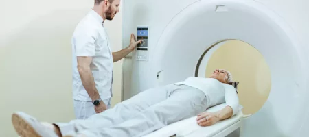
During the procedure, the patient lies on a flat surface on their back and is placed inside the machine. Complete stillness is required throughout the procedure, as even slight movement can blur the images. The procedure, which typically lasts 15–45 minutes, is painless. The machine produces loud noises while operating, so headphones or earplugs are provided. In some cases, a contrast agent may be administered intravenously to allow a more detailed examination. We manage the entire process for patient comfort, ensuring a safe and comfortable experience.
Types of MRI
With technological advancements, different types of MRI have emerged. Brain MRI, neck MRI, lumbar MRI, knee MRI, cardiac MRI, functional MRI, angiography MRI, and diffusion MRI are among the most well-known types. Sometimes a contrast MRI is preferred, while in other cases a non-contrast MRI is sufficient. The choice of method depends on the disease type and the area the doctor wants to examine. We select the most appropriate MRI type for each patient, ensuring the clearest and most reliable images.
What is the Purpose of MRI?
This method allows detailed examination of the internal structures of the human body. Brain vessel blockages, tumors, spinal and joint disorders, structural heart abnormalities, or masses in abdominal organs can be clearly detected through MRI. It also plays a crucial role in monitoring the treatment process and can be safely used before and after surgical interventions. Through MRI, we provide patients with the opportunity for early diagnosis and contribute to fast and accurate resolution of health issues.
When is MRI Needed?
MRI is one of the most reliable diagnostic methods for many health issues today. It is especially recommended for neurological symptoms such as prolonged headaches, memory loss, sudden vision loss, fainting, dizziness, balance problems, and suspected strokes. Additionally, severe pain or limited movement in the spine or joints, as well as post-traumatic injuries, are indications for MRI. If there is suspicion of tumors, masses, or vascular blockages in abdominal or thoracic organs, MRI is recommended for detailed evaluation. We use this method to accurately assess patient complaints and secure their health through early diagnosis.
Reasons for MRI
Doctors generally request MRI to clarify diagnosis, determine the severity of a disease, and plan treatment. It is used to examine brain vessels, detect spinal cord pressure, evaluate herniated discs, and diagnose muscle or ligament injuries. It is also preferred to visualize structural heart defects and investigate tumors or masses in internal organs like the liver, kidneys, or pancreas. MRI is valuable for pre-surgical planning and post-operative follow-up. Through this, we provide patients not only with diagnosis but also a safe guide throughout the treatment process.
Preparations Before MRI
Before the procedure, patients should not have metal accessories, jewelry, watches, belts, or credit cards that may affect the magnetic field. If the patient has implants such as a pacemaker, prosthesis, or cochlear implant, it must be reported to the doctor. Most MRI procedures do not require fasting, but for abdominal or pelvic imaging, doctors may request fasting. Wearing comfortable clothing facilitates the procedure. We provide detailed guidance before the procedure, ensuring a smooth and safe imaging experience.
Advantages of MRI
This method allows detailed imaging without exposing the body to radiation. Its greatest advantage is showing soft tissues, blood vessels, muscles, and nerves in fine detail. It provides high accuracy for evaluating the brain, spine, heart, joints, and internal organs. Contrast-enhanced MRI highlights vascular structures and possible tumors clearly. It also provides reliable information for pre-surgical planning and post-operative follow-up. We leverage these advantages to ensure accurate diagnosis and treatment for our patients.
Disadvantages of MRI
While MRI is a reliable method in many aspects, it has some disadvantages. The patient must remain still for an extended period, which can be challenging for children and claustrophobic individuals. The loud noise produced by the machine can be disturbing, so earplugs or headphones are used. MRI may not be suitable for patients with pacemakers, brain aneurysm clips, or metal implants. Rarely, the contrast agent may cause an allergic reaction. We evaluate these risks beforehand and ensure a safe and comfortable imaging experience for our patients.
Diseases Evaluated with MRI
MRI can be used to diagnose a wide range of diseases. It is preferred for neurological conditions such as brain vessel blockages, stroke, tumors, epilepsy, and multiple sclerosis. It clearly shows spinal and joint issues such as herniated discs, arthritis, and meniscus tears. Congenital heart anomalies, vascular narrowings, and myocarditis can also be evaluated. Masses, tumors, or inflammatory processes in internal organs such as the liver, kidneys, and pancreas are examined in detail. We use MRI not only for diagnosis but also for disease monitoring.
Organs Examined with MRI
Nearly all regions of the body can be evaluated with MRI. The brain, spine, neck, lumbar and back regions, heart and vessels, liver, kidneys, bile ducts, pancreas, uterus, and ovaries can be imaged in detail. Additionally, joints, muscles, ligaments, and vascular systems can be thoroughly examined. We use this broad evaluation capability to provide patients with a comprehensive assessment.
Types of MRI Imaging
With technological advancements, MRI applications have expanded. Brain MRI, neck MRI, lumbar MRI, cardiac MRI, angiography MRI, functional MRI, diffusion MRI, and spectral MRI are commonly performed. Whole-body MRI can scan multiple regions simultaneously. Both contrast and non-contrast options provide detailed information about organ structure and function. We select the appropriate MRI technique for each patient to ensure the most reliable results.
MRI Prices 2026
Prices vary depending on the region to be imaged, whether contrast is used, the technology of the device, and the location of the healthcare center. As of 2026, prices may differ at each center. The average MRI procedure may cost approximately 5,000–15,000 TL. For accurate pricing, patients can contact us for details. Our transparent pricing policy ensures that patients receive up-to-date and reliable information and affordable solutions.
Which Doctor Requests MRI?
MRI is an essential imaging method in diagnosing diseases. Many specialists may request MRI to clarify a diagnosis or assess disease severity. Neurologists, neurosurgeons, and orthopedists frequently use MRI for brain, nervous system, spine, and joint issues. Cardiologists prefer MRI for heart and vascular evaluation, while internal medicine specialists and oncologists use it for suspicious masses in internal organs. Gynecologists and obstetricians may use MRI to examine uterine and ovarian conditions or during pregnancy. We provide MRI services safely and comfortably for patients referred from various specialties, ensuring accurate diagnosis and smooth treatment.
Does MRI Hurt?
One of the most common patient concerns is whether MRI causes pain. The MRI procedure is completely painless. The patient lies on the machine table on their back and remains still throughout the scan. No incisions, needles, or surgical procedures are involved, except for contrast-enhanced MRI, which may involve a brief needle prick. Apart from that, no pain is felt during the procedure. The machine’s noise may be uncomfortable, but earplugs or headphones reduce this. We provide full support to minimize patient anxiety and ensure a comfortable and safe experience.
Difference Between CT and MRI
Both methods provide imaging of internal body structures, but there are important differences. CT uses X-rays and involves some radiation, making it advantageous for imaging air-filled organs like the lungs and bone structures. MRI, on the other hand, uses a powerful magnetic field and radio waves and does not involve radiation. MRI is superior for examining soft tissues, muscles, nerves, blood vessels, and sensitive organs like the brain. CT is faster, while MRI takes longer but offers higher detail. We determine the most appropriate method for each patient to ensure the most reliable diagnosis.
Does MRI Show Everything?
MRI is one of the most sensitive techniques for evaluating soft tissues. The brain, spinal cord, joints, muscles, ligaments, and internal organs can be clearly imaged. However, not everything can be detected solely with MRI. Small calcifications or some fine bone details may be better visualized with CT. MRI also does not provide biochemical information; it only shows anatomical structures. Therefore, it may need to be supplemented with blood tests or other imaging methods. Nevertheless, MRI is considered a gold standard for diagnosing many conditions. We combine MRI with other methods to ensure the most accurate diagnosis and guide treatment safely.
What to Do After MRI?
After the scan, patients often wonder about returning to daily life. Since MRI involves no radiation, patients can immediately resume normal activities after non-contrast scans. There is no pain or lasting discomfort. Some may experience mild stiffness or fatigue from lying still, which usually resolves quickly. If contrast was used, it is excreted by the kidneys, and drinking plenty of water is recommended. Temporary dizziness or nausea may occur in some patients but resolves on its own. No special diet or medication is required post-MRI. We provide detailed post-procedure guidance and answer any questions to support our patients throughout the process.
When Are MRI Results Available in Public Hospitals?
The time to prepare reports in public hospitals varies depending on the hospital’s workload. In busy urban centers, results may take several days, while in smaller centers it may be faster. On average, MRI results are available within 2–7 days. In emergency cases, reports can be expedited and provided the same day. Private clinics and advanced imaging centers often provide results within a few hours. We use technology to accelerate reporting and provide patients with reliable results without delay, ensuring timely treatment.
Which Diseases Can Be Detected with Spine MRI?
Spine MRI provides detailed imaging of the spine and surrounding tissues. It can reveal conditions causing back pain, such as herniated discs, disc slippage, spinal stenosis, tumors, infections, and spinal inflammation. It also evaluates blood supply to the spinal cord, muscle abnormalities, and congenital structural issues. Patients experiencing prolonged back pain, numbness, weakness, or limited mobility are often referred for this scan. MRI images help doctors determine the disease severity, progression, and treatment plan. We use advanced devices to ensure no detail is missed.
How Long Does a Closed MRI Take?
Closed MRI machines provide high-resolution images through a strong magnetic field. The duration varies depending on the region being scanned. An average closed MRI takes 15–45 minutes. Scanning a single area may take less time, while multiple regions like the brain, spine, or abdomen may take longer. Patients must remain still during the scan. Claustrophobic individuals may find it challenging, but earphones, music, and communication with staff help. Sedatives may be provided if necessary. We prioritize patient comfort and ensure safe and reliable results.
Can You Bathe After MRI?
After the imaging procedure, patients often ask if they can bathe. MRI does not involve incisions, needles, or external radiation, so daily activities, including bathing, can resume immediately. If contrast was used, doctors may recommend drinking fluids to help excrete it. Bathing is not restricted. We explain post-procedure instructions clearly and resolve any patient concerns.
Does MRI Affect the Lower Body?
Patients often wonder if MRI affects the lower body. MRI works with a strong magnetic field and radio waves, showing soft tissues in great detail. It does not capture dense bone structures as clearly as X-ray or CT, but provides extensive information about bone marrow, cartilage, ligaments, muscles, and surrounding soft tissues. It is commonly used for detailed evaluation of the lower back, knees, and hips. We guide patients on the appropriate device and scan type for their needs.
Does MRI Show Bones?
MRI plays an important role in imaging bone tissue. While X-rays show external bone structure, MRI provides detailed images of internal bone structures, bone marrow, cartilage, and joint surfaces. It is valuable for detecting tumors, inflammation, edema, or bone marrow disorders. X-rays are preferred for fractures, while MRI is better for soft tissue damage and bone marrow-related conditions. We offer both X-ray and MRI options and use them as needed.
CT or MRI: Which is Better?
The better method depends on the area to be examined. CT provides fast imaging of bones, lungs, and acute conditions. MRI offers superior resolution for the brain, spinal cord, muscles, joints, and soft tissues. Therefore, no single method is universally better. The doctor determines the most suitable scan based on patient complaints. We explain the advantages of each method and guide patients to make an informed choice.
Who Performs MRI?
The person performing MRI is called a “radiology technician” or “radiologic technologist.” They operate the device, position the patient, and ensure high-quality imaging. They communicate with the patient throughout the procedure to ensure safety and comfort. After images are obtained, radiologists analyze them and prepare the report. Thus, the technician conducts the scan, while the radiologist interprets and reports the results. We work with experienced technicians and specialist radiologists to ensure reliable evaluation of MRI results.


