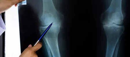
What is Knee MRI?
Knee MRI is a modern diagnostic method that uses magnetic resonance imaging to examine the knee joint and surrounding soft tissues in detail. Unlike methods such as X-ray or CT scan, it does not involve radiation. Thanks to powerful magnetic fields and radio waves, ligaments, cartilage structures, menisci, muscles, vessels, and bones in the knee joint can be clearly visualized. It is one of the most commonly used methods by doctors, especially in sports injuries, traumas, and chronic knee pain. We perform this advanced imaging technique for our patients in a safe, painless, and comfortable way, enabling them to reach an accurate diagnosis and treatment quickly.
How is a Knee MRI Performed?
During a knee MRI, the patient usually lies on their back, and the leg is stabilized. Some support devices may be used to prevent movement. For accurate imaging, the patient must remain still throughout the procedure. The process takes about 15 to 30 minutes. The machine may produce loud noises during the scan, so earplugs or headphones are used. In some cases, a contrast agent may be injected intravenously to provide a more detailed view of the tissues. We ensure comfort for our patients during the procedure and provide information before and after the scan to make the process smooth.
Why is a Knee MRI Requested?
Doctors may request a knee MRI to determine the cause of complaints in the knee joint. This method is particularly useful for examining meniscus tears, anterior cruciate ligament (ACL) injuries, cartilage damage, edema, and fluid accumulation around the knee. It is also preferred for chronic knee pain, joint stiffness, movement limitations, unexplained swelling, and suspected tumors. Physicians frequently rely on this method for accurate diagnosis. We provide detailed results with knee MRI to reveal the underlying causes of our patients’ complaints clearly.
In Which Cases is a Knee MRI Performed?
Knee MRI is commonly required in cases of trauma to the knee, injuries after falls, or strain caused by sports activities. It is also used to diagnose meniscus injuries, ligament damage, osteoarthritis, rheumatic diseases, increased joint fluid, cartilage wear, and bone marrow edema. Knee MRI is also crucial in pre-surgical evaluations, allowing the joint structure to be assessed in detail before surgery. We perform this procedure to provide reliable data both before surgery and during treatment.
Where is a Knee MRI Performed?
Knee MRI is performed in equipped imaging centers and hospitals. These centers have advanced MRI machines, and the scan is carried out by specialized radiology technicians. The images obtained are then reported by a radiology specialist. Today, many private healthcare facilities and state hospitals offer knee MRI services. We perform knee MRI using advanced technology to provide our patients with safe and high-quality service.
Is Knee MRI Harmful?
Since knee MRI is performed using magnetic resonance imaging, it is not considered harmful because it does not involve radiation. Unlike X-ray or CT scans, no radiation is used. Instead, powerful magnetic fields and radio waves provide detailed imaging of tissues. Therefore, patients do not experience any harmful effects during or after the procedure. It has been safely applied for many years. However, caution is required in patients with pacemakers, brain stimulators, metal implants, or prosthetics. We inform our patients in advance and take necessary safety precautions before the procedure.
What are the Risks of Knee MRI?
For most patients, knee MRI does not pose any significant risks. However, certain situations require caution. For example, people with metal implants, prosthetics, or stents sensitive to magnetic fields may face risks. The magnetic field may cause these materials to move or malfunction. Additionally, when a contrast agent is required, caution is needed in patients with impaired kidney function. Very rarely, allergic reactions may occur due to contrast agents. We carefully review our patients’ medical history before the procedure to minimize these risks and ensure a safe process.
What are the Advantages of Knee MRI?
MRI is one of the most effective methods for detailed imaging of the knee joint. Its main advantage is its ability to display soft tissues, cartilage, menisci, and ligaments in high resolution. This allows accurate diagnosis without surgery. The procedure is painless, radiation-free, and completed quickly. Patients can return to normal life immediately after the scan. It is also highly beneficial in surgical planning. With this method, we help our patients reach accurate diagnoses faster and simplify the treatment process.
How to Prepare for a Knee MRI?
Knee MRI does not require special preparation. However, patients should not wear clothing or carry items with metal components, such as jewelry, watches, or credit cards, which may be affected by magnetic fields. In some cases, short fasting may be required if a contrast agent is used. Wearing comfortable clothing improves comfort during the procedure. Remaining still is essential for clear images. We inform all our patients beforehand, make preparations easier, and ensure the scan is performed smoothly.
How Long Does a Knee MRI Take?
Knee MRI, used to examine the internal structure of the knee joint, usually takes about 20 to 40 minutes. The duration depends on the technology of the machine, whether contrast is used, and whether the patient remains still. For some patients, it may take longer if remaining motionless is difficult. During the procedure, the patient lies on a comfortable table and is placed inside the scanner. The process is painless. We provide support to make the procedure as comfortable as possible, even guiding patients on breathing if needed.
How Long Does it Take to Get Knee MRI Results?
After the scan, the images are reviewed by a radiologist. On average, results are available within a few hours to one day. In urgent cases, reports can be prepared much faster. Preparing a detailed report requires the doctor to carefully analyze the images and compare them with other findings if necessary. Using advanced technology, we ensure results are delivered quickly, reliably, and clearly to our patients, allowing treatment to begin promptly.
What Can Be Seen in a Knee MRI?
This imaging method allows detailed visualization of menisci, ligaments, cartilage, joint fluid, and surrounding soft tissues in the knee. Trauma-related injuries, ligament tears, meniscus tears, cartilage damage, inflammation, and cysts can be clearly identified. It is particularly reliable in diagnosing knee injuries commonly seen in athletes. With this examination, we provide accurate diagnoses and manage treatment more effectively.
Which Diseases Can Be Diagnosed with a Knee MRI?
Diseases commonly diagnosed with knee MRI include meniscus tears, anterior and posterior cruciate ligament injuries, joint inflammations, bone marrow edema, cysts, joint effusion, tumors, and cartilage damage. Changes caused by rheumatic diseases in the joints can also be clearly observed. It is particularly useful for people with chronic knee pain or recurrent trauma-related problems. Therefore, it is one of the most reliable methods both for diagnosis and treatment planning.
Knee MRI Prices 2026
Prices vary depending on the technology of the machine, whether contrast is used, the location of the healthcare facility, and any additional services required by the patient. If contrast or high-resolution devices are used for detailed imaging, the cost may range from approximately 7,000 to 15,000 TL or more.
With our transparent pricing policy, we provide patients with clear and up-to-date information, ensuring affordable yet high-quality services. In this way, we deliver both reliable and accessible healthcare.
Do You Need to Remove Clothes for a Knee MRI?
Since MRI scanners use a strong magnetic field, clothing with metal components must be removed. If pants or tops contain zippers, buttons, or accessories, they must be changed. In most cases, patients are provided with a special hospital gown. This gown ensures comfort and prevents anything from affecting image quality. We inform patients about what clothing needs to be removed before the procedure to ensure safety and a smooth process.
What to Do After an MRI?
No special rest is required after an MRI. Patients can return to daily activities the same day. If a contrast agent was used, drinking plenty of water is recommended to help eliminate the contrast through the kidneys. Some patients may feel mild fatigue or dizziness, but this usually resolves quickly. We inform our patients after the procedure and, if necessary, follow up with a check-in call to prevent possible side effects and ensure a safe process.
Can MRI Show Everything?
MRI is extremely effective for examining soft tissues. Muscles, ligaments, cartilage, menisci, blood vessels, and joint fluid can be clearly seen. However, some minor calcifications or very fine bone structures may be better visualized with other imaging methods. Therefore, although MRI may not show everything, it is highly effective in diagnosing many conditions and problems. We use this method to achieve highly accurate results and plan treatments more effectively.
What is Knee Effusion?
Effusion is the swelling and pressure sensation caused by excess fluid accumulation in the joint area. Knee effusion often occurs due to trauma, strain, meniscus tears, ligament injuries, or rheumatic diseases. This condition causes pain, restricted movement, and discomfort. MRI helps identify the cause and severity of effusion clearly, enabling accurate treatment planning. In such cases, we provide early diagnosis and ensure our patients have a comfortable and faster recovery process.
Can You Drink Water Before an MRI?
Generally, drinking water before an MRI is not a problem. In fact, for contrast MRIs, light fluid intake beforehand may be helpful. However, drinking excessive amounts of water that fill the stomach is not recommended. For abdominal MRIs, fasting may sometimes be required depending on the doctor’s instructions. For knee MRIs, there is no such requirement. We provide necessary instructions to our patients before the procedure to ensure proper preparation.
Do You Have to Remove a Bra During an MRI?
MRI scanners generate a strong magnetic field, so all clothing or accessories containing metal must be removed. Bras with metal wires or clasps can interfere with image quality and pose safety risks. Therefore, patients are usually asked to remove them. If the bra does not contain metal, removal may not always be necessary, but many centers require it as standard practice. We provide our patients with clear instructions before the procedure and supply gowns to ensure comfort.
Who Cannot Have an MRI?
Like all imaging methods, MRI has some limitations. Patients with pacemakers, internal metal prosthetics, or brain vessel clips should not undergo MRI. Additionally, patients with severe claustrophobia may not tolerate the procedure. MRI is also generally avoided in the first trimester of pregnancy unless absolutely necessary. We collect detailed health information from our patients beforehand to ensure MRI can be performed safely.
Do You Have to Fast Before an MRI?
Not all MRIs require fasting. For example, MRIs of the knee, brain, or spine do not require fasting. However, abdominal and pelvic MRIs may require fasting for clearer images. This is because a full stomach and intestines can distort the images. Fasting helps organs appear more clearly. We inform patients individually whether fasting is needed to ensure optimal results.
Why Does the MRI Machine Make Noise?
During MRI, the coils inside the scanner rapidly change current within the strong magnetic field. This creates vibrations and loud sounds. The knocking or tapping noises heard during the scan are caused by this process. Most centers provide earplugs or headphones to minimize discomfort. The noise is a normal part of the scanner’s operation and is not dangerous. We ensure our patients’ comfort by providing necessary equipment during the scan.
What Helps with Knee Osteoarthritis?
Knee osteoarthritis occurs when the cartilage covering joint surfaces wears down over time, reducing mobility and causing pain. Lifestyle changes play an important role in managing this condition. Regular exercise maintains joint mobility, strengthens muscles, and reduces pain. Weight loss reduces stress on the knee joint and slows progression. Additionally, doctor-prescribed pain relievers and anti-inflammatory medications provide relief. Physiotherapy, intra-articular injections, and PRP therapy are also commonly used methods in recent years. We provide our patients with tailored treatment plans and closely monitor their progress.
How to Tell if You Have a Meniscus Injury?
The menisci in the knee joint can tear due to sudden movements, sports injuries, or trauma. Patients with a meniscus tear often feel sharp pain when twisting the knee. Locking, giving way, and swelling of the knee are common symptoms. Pain may also worsen when climbing stairs or standing up. MRI is required for an accurate diagnosis. We use both clinical examination and imaging methods to make a correct diagnosis in such cases.
How to Tell if You Have Knee Osteoarthritis?
Osteoarthritis is usually a slowly progressing condition that becomes noticeable over years. Stiffness in the knees, difficulty climbing stairs, pain after prolonged walking, and cracking sounds during movement are typical signs. Morning stiffness or pain after prolonged inactivity is also common. In the early stages, symptoms may be mild but worsen over time. X-rays and MRIs can clearly show the degree of osteoarthritis. We perform detailed evaluations for patients with these complaints and provide personalized treatment plans.
How to Tell if the Kneecap is Dislocated?
Patellar dislocation usually occurs suddenly after trauma. Patients experience severe pain, visible deformity, and loss of mobility. The kneecap often shifts to the side, causing obvious shape changes. Bearing weight becomes nearly impossible. Emergency intervention is necessary, followed by imaging studies to assess ligaments and cartilage. We respond quickly in such emergencies to ensure safe treatment management.
Is X-ray Used for Knee Pain?
X-ray is one of the most common diagnostic tools for knee pain. It is especially useful for detecting fractures, dislocations, osteoarthritis, or deformities in bone structures. However, X-rays may not be sufficient for soft tissue injuries, in which case MRI is needed. X-ray is quick, accessible, and practical. For patients with knee pain, we first perform a detailed examination and then decide which imaging method is appropriate, ensuring accurate diagnosis and effective treatment planning.
When Are Non-Contrast MRI Results Available?
For MRIs without contrast, results are generally available faster. After the scan, the radiologist reviews the images in detail. Reports are usually ready within a few hours to one day. In urgent cases, results may be provided much faster. Since non-contrast MRI does not require additional preparation, the process is more practical. We use advanced technology to deliver results quickly and reliably to our patients.
What Can Be Seen in a Leg MRI?
MRI of the lower extremities provides detailed imaging of muscles, vessels, joints, ligaments, and bones. Injuries from trauma, muscle tears, vascular blockages, tumors, infections, and cysts can be clearly detected. It also provides important information for diagnosing circulation disorders and musculoskeletal diseases. This allows detailed evaluation without the need for surgery. With leg MRI, we accurately identify the cause of patients’ complaints and provide effective treatment processes.
What Causes Knee Pain?
Knee pain can result from many different causes. The most common are meniscus tears, ligament injuries, osteoarthritis, rheumatic diseases, excess weight, sports injuries, and trauma. Patellar dislocations, increased joint fluid, and cartilage damage can also cause pain. Sometimes pain may result from short-term strain, but if it becomes chronic, further investigation is needed. We provide detailed examinations and appropriate imaging methods to identify the exact cause in our patients.
Can MRI Be Performed with Knee Implants?
Metal implants, plates, or prosthetics in the body require careful consideration for MRI. Most modern plates and orthopedic implants are MRI-compatible, meaning the scan can be performed safely. However, older types of metal may pose risks. Additionally, the presence of metal in the scanned area may distort images. Therefore, patients must inform their doctor beforehand. We ensure necessary checks are done to perform imaging safely.
Can You Wear Jeans During an MRI?
Jeans with metal buttons, zippers, or accessories are not suitable for MRI. The magnetic field may affect metal parts and distort images. Therefore, patients are usually asked to change clothes before the procedure. Most centers provide special gowns. We provide suitable clothing for our patients to ensure comfort and smooth imaging.
Can Meniscus Injuries Be Seen on MRI?
Yes, meniscus tears or damage can be clearly diagnosed with MRI. The structure of the meniscus, as well as the size and location of the tear, can be visualized in detail. For this reason, MRI is the most commonly used and reliable method for diagnosing meniscus problems. We use this method to provide accurate diagnoses and create personalized treatment plans for our patients.
Can You Shower After an MRI?
If no contrast agent is used, showering after MRI is safe. Even if contrast is used, showering the same day is usually allowed. However, the injection site may be slightly red or sensitive, so it should be cleaned gently. We provide our patients with the necessary information after the procedure to ensure they safely return to their daily routine.


