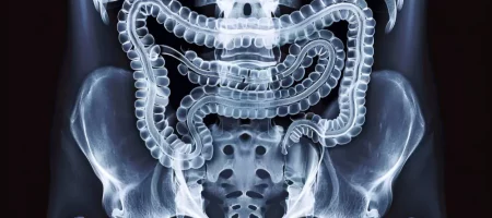
What is Enteroclysis?
There is a special imaging method that allows detailed examination of the small intestines. This method was developed to evaluate the structure, movements, and any abnormal formations of the intestines. It is particularly important in digestive system disorders, especially when unexplained abdominal pain, chronic diarrhea, or suspected small bowel obstruction is present. Using this technique, the intestines are filled with a contrast agent, and images are taken via X-ray or CT devices. With this method, we speed up diagnosis in our patients and ensure that the correct treatment plans are made.
How is Enteroclysis Performed?
Before the procedure, preparation is made to empty the patient’s intestines. This usually requires fasting for a certain period and, in some cases, taking medication for bowel cleansing. During the imaging, a contrast agent is administered directly into the small intestine through a thin tube. Then, imaging is performed using the device. This allows the structural and functional condition of the intestines to be clearly observed. During the procedure, the patient should remain still and follow the instructions of the technician and radiologist. We prioritize the comfort of our patients and ensure that the imaging process is as comfortable as possible.
How Many Types of Enteroclysis Are There?
There are different enteroclysis methods depending on the application. The most well-known are conventional enteroclysis and CT enteroclysis. In the conventional method, images are taken using X-ray after administering a contrast agent. CT enteroclysis, on the other hand, is performed with computed tomography and allows much more detailed imaging. Nowadays, a method called MR enteroclysis is also used, in which the small intestines can be evaluated more comprehensively using an MRI device. We select the most suitable method according to the patient’s condition, ensuring the correct diagnosis is made quickly.
When is Enteroclysis Performed?
This imaging technique is used when digestive system complaints cannot be explained by conventional methods. It is particularly important for chronic bowel diseases such as Crohn’s disease, small intestine tumors, unexplained abdominal pain, and suspected bowel obstruction. Additionally, it may be used in cases of diarrhea, anemia, and weight loss. Early diagnosis allows many diseases to be treated more effectively. With this method, we provide reliable results to our patients and contribute to proper treatment planning.
Contraindications of Enteroclysis
There are certain situations where medical procedures cannot be applied, and enteroclysis is among them. This method is not performed in cases of complete bowel obstruction, acute abdomen, suspected bowel perforation, or severe infection. Radiation-containing methods are avoided in pregnant patients. If the patient has a history of allergies, sensitivity to the contrast agent is evaluated. The doctor plans the procedure taking all these risks into account. We also inform our patients in detail before the procedure, ensuring a safe process.
Diseases Where Enteroclysis is Used
Some digestive system disorders require more detailed examination. This method plays a significant role in chronic abdominal pain, unexplained diarrhea, and suspected small bowel strictures or obstructions. It facilitates the diagnosis of inflammatory bowel diseases, especially Crohn’s disease. It is also preferred for investigating tumors, polyps, or masses in the small intestine. It is highly effective in monitoring disease progression and changes in the intestinal wall. With this method, we provide early diagnosis opportunities for our patients and manage the treatment process more safely.
Where is Enteroclysis Used?
Medical imaging centers and advanced hospitals can perform this procedure because it requires specialized equipment and experienced specialists. It is one of the most effective methods to examine intestinal movements and structure. It can be performed using conventional X-ray devices as well as with CT or MRI. It is a technique used especially when other imaging methods are insufficient for small intestine issues. With our modern devices, we perform this examination safely and provide the most accurate results to our patients.
Who Can Undergo Enteroclysis?
It can be applied to individuals with persistent abdominal pain, unexplained anemia, unexplained weight loss, chronic diarrhea, or constipation. It is also preferred for patients suspected of having intestinal strictures or adhesions. It is useful for monitoring the course of chronic diseases such as Crohn’s disease. However, the procedure is not performed in certain high-risk groups, such as patients with complete bowel obstruction or severe infection. Patient safety is always a priority. We carefully evaluate our patients’ conditions and perform the procedure when appropriate.
Preparations Before Enteroclysis
For the procedure to be performed effectively, the intestines must be empty. Therefore, patients are asked not to eat for a certain period before imaging. In some cases, medications may be used to cleanse the bowel. The patient’s previous medical history, medications, and allergy history should also be communicated to the doctor. Wearing comfortable clothing and following the doctor’s instructions is important. We provide detailed guidance to our patients before the procedure and make the preparation process easier.
Enteroclysis Prices 2026
The prices of imaging methods vary depending on the device used, the type of procedure (conventional, CT, or MR enteroclysis), and the facility. As an approximate estimate, you can budget around 7,000–14,000 TL, leaving some margin.
The contrast agent used during imaging also affects the cost. Contact us for the most up-to-date and accurate information. We always prioritize patient satisfaction and aim to provide high-quality services under suitable conditions.
Is There a Diet Restriction Before Enteroclysis?
Before any bowel imaging procedure, it is essential that the intestines are clean. Therefore, patients are usually asked to follow a food and drink restriction for a certain period. The day before the procedure, light foods should be preferred, avoiding gas-producing foods. Sometimes, the doctor may recommend a laxative or enema to ensure the bowel is completely empty. Drinking water is usually allowed, as keeping the intestines hydrated helps the contrast agent distribute evenly. We provide detailed instructions to our patients about which foods they can consume, ensuring high-quality imaging.
How is CT Enteroclysis Performed?
In this procedure performed with computed tomography, the contrast agent is administered into the small intestine via a special tube. Then, the CT device is used to obtain detailed images of the intestines. This method provides much more detailed information than conventional enteroclysis and can show even minor changes in the intestinal wall. During the procedure, the patient lies inside the device, and the imaging usually takes a short time. Necessary measures are taken to maintain patient comfort throughout the procedure. We perform CT enteroclysis safely with modern devices, providing our patients with highly accurate results.
What Does Enterography Mean?
In medicine, enterography is a special radiological method performed to image the small intestines. Unlike conventional enteroclysis, the contrast agent is administered orally, and it is allowed to progress naturally with intestinal movement. Then, the small intestine is imaged in detail with a CT or MRI device. It is particularly effective in monitoring diseases such as Crohn’s disease that affect the small intestines. It is also frequently used to detect strictures, polyps, or masses. We also offer enterography to our patients, applying the most appropriate imaging techniques for different needs.
How is a Barium Small Bowel X-ray Performed?
In this method used to evaluate the structure and function of the intestines, the contrast agent plays a crucial role. Before the procedure, bowel cleansing is performed, and then the contrast agent is administered. This agent fills the intestines and allows them to be clearly seen on X-ray. During the procedure, the patient is positioned in various ways to capture the intestines from different angles. The process can be completed in approximately 20–40 minutes. After the procedure, patients can return to daily life, but it is recommended to drink plenty of fluids to help flush out the contrast agent. We perform these barium X-rays safely, assisting our patients in obtaining an accurate diagnosis.
How is a Small Bowel X-ray Taken?
The small intestine, one of the longest parts of the digestive system, sometimes needs detailed imaging for certain conditions. One of the methods used is a barium small bowel X-ray. Before the procedure, patients are usually placed on a restricted diet to empty the intestines. In some cases, bowel-cleansing medications may also be used.
During imaging, a contrast agent is administered to make the intestines more visible on X-ray. This agent usually contains barium sulfate and helps produce detailed images of the intestinal wall. The patient is asked to lie in different positions to capture the entire small intestine. The procedure usually takes 30–60 minutes. We apply these imaging methods with the most modern devices, providing our patients with reliable results.
Why is Small Bowel MRI Taken?
For some conditions, conventional X-ray or CT may not be sufficient. At this point, magnetic resonance imaging (MRI) comes into play. Small bowel MRI is preferred in cases of Crohn’s disease, intestinal inflammation, tumors, or small bowel strictures. This method allows detailed assessment of both the intestinal wall thickness and surrounding tissues.
Since it does not involve radiation, it is safe for children and young patients as well. Early diagnosis through MRI can make a significant difference in the treatment process. We perform small bowel MRIs with our expert team, providing the highest contribution to the diagnostic process.
What are Enterocyte Cells?
Enterocytes are one of the most important cells responsible for digestion and absorption in the intestines. Located on the surface of the small intestine, these cells allow nutrients from food to enter the bloodstream. They play an active role in breaking down and absorbing carbohydrates, fats, and proteins. They also have an important function in the immune system.
If enterocytes do not function properly, problems such as diarrhea, nutrient absorption disorders, and weight loss may occur. Diseases like celiac disease and Crohn’s disease can damage these cells. We carefully perform all diagnostic and follow-up procedures related to small bowel health, providing our patients with the most accurate treatment plans.
What are Small Bowel Diseases?
Many diseases can develop in the small intestine, an important part of the digestive system. The most common problems include Crohn’s disease, celiac disease, small bowel tumors, diverticula, absorption disorders, and intestinal infections. Some of these diseases are chronic and require long-term follow-up.
Symptoms include abdominal pain, diarrhea, anemia, weight loss, bloating, and digestive disorders. Early diagnosis increases treatment success. With modern imaging methods and expert doctors, we provide the most reliable services in the diagnosis and follow-up of these diseases.
Why is Small Bowel Transit X-ray Taken?
Small bowel transit X-ray evaluates intestinal motility and transit speed. The patient orally consumes a contrast agent, and its progress through the intestines is monitored with X-ray images taken at regular intervals. This helps determine if there is a slowdown, acceleration, or obstruction in intestinal movement.
It is particularly preferred in chronic constipation, abdominal pain, suspected bowel obstruction, and absorption disorders. No surgical intervention is required during the procedure; it is completely non-invasive. We perform small bowel transit X-rays safely, providing our patients with fast and accurate diagnostic results.
What is an Intestinal X-ray?
An intestinal X-ray is a method used to diagnose digestive system diseases. It plays an important role in evaluating both the small and large intestine. The procedure usually involves a contrast agent, which coats the inner surface of the intestines and makes them clearly visible under X-ray. This allows strictures, dilations, polyps, tumors, or inflammation to be detected easily.
Intestinal X-ray enables early detection of many conditions that are difficult to diagnose through standard examination. The patient is positioned in different ways during the procedure, allowing detailed imaging of all intestinal regions. We perform these X-rays safely with modern devices, providing the most accurate diagnostic data to our patients.
Can Colon Cancer be Detected with CT?
Computed tomography (CT) is very effective in evaluating the structure of the intestines and colon. In cases of colon cancer, CT can detect masses, wall thickening, or suspicious lesions in the colon. Especially with contrast-enhanced CT, the internal structure of the bowel can be examined in detail.
CT alone may not be sufficient for colon cancer diagnosis; it is generally evaluated alongside colonoscopy. However, CT is crucial in assessing the spread and staging of cancer to surrounding tissues. We perform necessary imaging for patients suspected of colon cancer, contributing to early diagnosis.
What is Crohn’s Disease?
Crohn’s disease is an inflammatory bowel disease affecting the intestines. It is a chronic condition that can occur in any part of the digestive system but is most commonly seen in the small intestine and the terminal part of the large intestine. It causes inflammation in the intestinal wall and can lead to strictures, fistulas, or adhesions over time.
Symptoms include abdominal pain, diarrhea, weight loss, anemia, fatigue, and fever. The disease usually flares up and then calms down periodically. Although the exact cause is unknown, the immune system, genetic factors, and environmental influences play a role. We use the latest imaging methods in diagnosing Crohn’s disease and provide long-term follow-up and treatment support.
How Long Does Enterography Take?
Enterography, used for detailed imaging of the small intestines, usually involves orally administering a contrast agent, which is then imaged with CT or MRI during its passage through the intestines. With preparations included, the procedure takes approximately 30 to 60 minutes.
The duration may vary depending on the patient’s intestinal condition, contrast agent, and imaging device used. Sometimes patients may need to wait for clearer images. Patients can return to daily life immediately after the procedure. We perform enterography comfortably, ensuring reliable results.
What are the Symptoms of Crohn’s Disease?
Crohn’s disease, one of the most important chronic inflammatory bowel diseases, presents symptoms that vary from person to person. The disease usually manifests with intestinal inflammation, strictures, or ulcers. Common symptoms include abdominal pain, diarrhea, loss of appetite, and weight loss. Some patients may also experience anemia, fatigue, fever, blood in stool, and mouth ulcers.
Symptoms can worsen or subside periodically. Therefore, persistent abdominal pain or diarrhea requires advanced diagnostic procedures. We use the latest imaging and laboratory methods for early diagnosis of Crohn’s disease and provide reliable solutions to our patients.
How is Small Bowel Cancer Diagnosed?
Although rare, small bowel cancer can have serious consequences and requires detailed diagnostic methods. Tumor or polyp presence is investigated through colonoscopy and endoscopy. Additionally, CT, MRI, and enteroclysis are used to examine the intestinal structure in detail.
If cancer is suspected, a biopsy is taken to confirm the diagnosis. Early detection increases the chances of successful treatment. Therefore, patients with persistent symptoms should undergo diagnostic procedures without delay. We perform all necessary tests to ensure accurate diagnosis for our patients.
Can Crohn’s Disease be Detected with MRI?
MRI is one of the most reliable methods for diagnosing Crohn’s disease today. Especially with MR enterography, the thickness of the intestinal wall, level of inflammation, fistulas, or abscesses can be seen in detail. MRI allows precise assessment of disease extent and severity.
Its non-radiation feature is a major advantage, making it suitable for long-term monitoring of chronic diseases like Crohn’s. We safely perform MRI imaging in patients with Crohn’s disease, providing important data for treatment planning.
Why is MR Enterography Performed?
MR enterography is used for diagnosing intestinal diseases. In this procedure, a contrast agent is taken orally to allow natural movement of the intestines, and detailed images are obtained with an MRI device. It is particularly preferred in cases of Crohn’s disease, ulcerative colitis, small bowel tumors, and strictures.
MR enterography allows evaluation of both changes in the intestinal wall and surrounding tissues. Being non-invasive and safe, it can also be used in children and young patients. We perform MR enterography with modern devices, providing early diagnosis and accurate follow-up for our patients.
What is Ileus?
Ileus refers to bowel obstruction, a serious condition requiring urgent intervention. In this disease, bowel movements stop or slow down, preventing the passage of food. Symptoms include severe abdominal pain, vomiting, gas, and inability to pass stool.
Ileus can be caused by mechanical factors (tumor, adhesions, hernia) or motility disorders. If untreated, it can lead to severe complications such as bowel perforation. We perform urgent imaging and laboratory examinations in suspected ileus cases, initiating fast and effective treatment.
What is Colon MRI?
Colon MRI is a method used to examine the structure of the large intestine. This imaging technique evaluates the colon wall and surrounding tissues without radiation using magnetic resonance technology. It is particularly preferred for diagnosing colon cancer, polyps, inflammatory bowel disease, and strictures.
Colon MRI provides a comfortable examination and detailed results. Using contrast agents, the inner surface of the colon can be visualized more clearly. We perform colon MRI scans with our expert team, offering the most reliable results to our patients.


