MRI, or magnetic resonance imaging, is an imaging method that uses strong magnetic fields and radio waves. Its main advantages are that it does not contain radiation, provides high-resolution images, and shows soft tissues in detail.
Scan Process: The patient is placed inside a magnetic tunnel-shaped device. It is essential not to move during the scan. The duration varies between 15 and 45 minutes depending on the region being examined.
Advantages include being radiation-free, providing detailed information about tumors, hernias, brain and spinal diseases. In addition, since it clearly shows soft tissues, it plays a critical role in surgical planning.
Possible Risks: MRI is not entirely harmless. It can be challenging for people with claustrophobia. It may not be suitable for those with pacemakers, metal implants, or prostheses. When contrast agents are used, allergic reactions may rarely occur.
In conclusion, MRI is one of the most reliable and effective imaging methods in modern medicine. When used correctly, it greatly facilitates both diagnosis and treatment planning.
Imaging methods used in medicine play a very important role in the early diagnosis of diseases and in treatment planning. Techniques such as X-ray, ultrasound, CT, and MRI allow detailed examination of the internal structure of the body. Among these, one of the most advanced and detailed methods is MRI (Magnetic Resonance Imaging). MRI stands out especially for its ability to show soft tissues and critical areas such as the brain and spinal cord in great detail.
So what is MRI, how is it performed, what are its advantages, and what possible risks does it have?
What is MRI?
MRI is an imaging technique that creates detailed images of the body using strong magnetic fields and radio waves. Since X-rays are not used, it does not involve radiation. With this feature, it is safely preferred in many diseases.
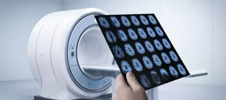
The MRI machine is designed as a large ring or tunnel. The patient lies inside this device and must remain completely still during the procedure.
In Which Cases is MRI Used?
MRI has a very wide range of applications.
- Brain and nervous system: Tumors, vascular blockages, multiple sclerosis
- Spine and joints: Lumbar hernia, cervical hernia, disc displacements
- Muscles and connective tissue: Sports injuries, tears, inflammations
- Internal organs: Masses in the liver, kidneys, pancreas, and uterus
- Vascular structure: Detailed examination of blood vessels with MR angiography

How Does the MRI Scan Process Work?
- Preparation
The patient is asked to remove all metal objects. Items such as necklaces, earrings, watches, phones, and credit cards may be damaged by the device’s magnetic field. - Position
The patient lies on the table and is placed inside the device. The position is adjusted according to the area to be examined. - Scan
During the scan, the device makes loud noises. Therefore, the patient is given headphones or earplugs. The scan usually lasts between 15 and 45 minutes. - Contrast MRI
In some cases, a contrast agent is injected intravenously. This makes blood vessels and tissues appear clearer. - Result
The obtained images are evaluated and reported by a radiology specialist.
Advantages of MRI
- Does not contain radiation: Unlike CT, X-rays are not used.
- Shows soft tissues clearly: Tumors, inflammations, and tissue damage can be easily detected.
- Versatile use: Can be applied to the brain, heart, joints, and internal organs.
- High resolution: Details not visible with other methods can be revealed with MRI.
- Guide in surgical planning: Provides critical information especially before brain and spinal surgeries.
Possible Risks and Disadvantages of MRI
- Claustrophobia: Its structure can be challenging for people with fear of closed spaces.
- Metal implants: Pacemakers, surgical clips, or metal prostheses can prevent MRI scans.
- Contrast agent: Although rare, allergic reactions may occur. It should be used with caution in people with kidney failure.
- Long scan time: Especially for children and restless patients, staying still can be difficult.
Precautions Before MRI
- All metal items such as jewelry, watches, and phones must be removed
- If there is a pacemaker or metal implant, the doctor must be informed
- If contrast imaging is to be performed, kidney function tests should be evaluated beforehand
- Patients should remain still during the scan
Difference Between MRI and Other Methods
- X-ray: Shows bone structures well but provides limited soft tissue images.
- CT (Computed Tomography): Very effective in imaging bones and lungs, but involves radiation.
- Ultrasound: A safe and practical method, but image quality may be limited.
- MRI: Shows soft tissues in the clearest way and does not involve radiation.
Since each method has different advantages, doctors usually choose the most suitable imaging method for the patient.
How Are MRI Results Interpreted?
MRI results are reported by a radiology specialist. However, for the report to be turned into appropriate treatment for the patient, it must be evaluated by the relevant physician. MRI only assists in diagnosis. A definitive diagnosis and treatment plan is always made together with a clinical examination.
Conclusion
MRI is one of the most reliable and advanced imaging methods in modern medicine. Thanks to its radiation-free nature, high-resolution imaging, and wide range of use, it is the first choice in many diseases.
Although it has some risks and disadvantages, when used under the right conditions, it provides great convenience in early diagnosis and treatment planning.
Having regular check-ups and undergoing appropriate imaging methods when necessary increases quality of life and helps prevent possible serious diseases. MRI is one of the most valuable diagnostic tools in this process.


