Modern imaging methods in medicine are indispensable in the diagnosis of diseases. MRI, CT, and ultrasound are methods that work with different techniques and serve different purposes.
MRI (Magnetic Resonance Imaging): The best method for visualizing soft tissues. It is preferred for detailed examination of the brain, spine, joints, and internal organs. It does not contain radiation. It is especially the first choice for brain tumors, herniated discs, or connective tissue injuries.
CT (Computed Tomography): Cross-sectional images are obtained using X-rays. It is very effective in evaluating the lungs, coronary arteries, internal bleeding, or fractures after trauma. Thanks to its rapid results, it is often used in emergencies.
Ultrasound: A method that works with sound waves and is harmless. It is preferred for the evaluation of abdominal organs, the thyroid gland, or pregnancy monitoring. It also provides guidance during biopsies.
Which of these three methods will be used depends on the patient’s complaints, the doctor’s preliminary diagnosis, and the organ to be examined. In some cases, the methods can also be used complementarily.
In conclusion, each imaging method has its own unique advantages. Using the right method at the right time makes a big difference in the early diagnosis of diseases.
With the advancement of technology in the medical world, the diagnosis and treatment processes of diseases have become much faster and more reliable. Especially imaging methods provide doctors with great advantages in making accurate diagnoses. MRI, CT, and ultrasound are the three most commonly used imaging techniques. However, most people do not know the differences between these methods, when they are used, and why.
In this article, detailed information about MRI, CT, and ultrasound will be provided, explaining in which situations they are preferred and their advantages.
The Role of Imaging Methods in Medicine
Imaging techniques allow the inside of the human body to be seen non-invasively. In this way, early diagnosis is ensured and the treatment process can be planned more accurately. Today, a single method is usually not sufficient in the diagnosis of diseases. Which imaging technique will be used depends on the patient’s complaints, the doctor’s preliminary diagnosis, and the organ to be examined.

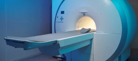
MRI (Magnetic Resonance Imaging)
MRI is a method that uses a strong magnetic field and radio waves to produce detailed images of the body. It does not contain radiation. For this reason, it is considered a safe method.
Uses of MRI
- Evaluation of brain tumors, vascular diseases, and strokes
- Diagnosis of spinal and lumbar hernias
- Examination of joint, ligament, and muscle injuries
- Detection of tumors or inflammations in internal organs
- Contrast imaging of vascular structures
Advantages of MRI
- Shows soft tissues very clearly.
- Does not contain radiation.
- High-resolution images from different angles can be obtained.
Disadvantages
- The scan time can be long.
- Challenging for those with claustrophobia.
- Limited use in people with metal implants.
CT (Computed Tomography)
Computed tomography provides cross-sectional images using X-rays. It is known for its ability to scan the entire body in a short time.
Uses of CT
- Diagnosis of lung diseases, pneumonia, or tumors
- Evaluation of coronary arteries (coronary CT angiography)
- Examination of internal bleeding and fractures after trauma
- Monitoring the spread of tumors
- Rapid diagnosis in emergency situations
Advantages of CT
- Provides results in a very short time.
- Shows bone structures clearly.
- Plays a vital role in emergency cases.
Disadvantages
- Contains radiation.
- Generally not preferred in pregnant women.
Ultrasound
Ultrasound is a method that visualizes organs using sound waves. It does not contain radiation and is considered very safe.
Uses of Ultrasound
- Pregnancy monitoring and fetal development
- Examination of organs such as the liver, kidneys, gallbladder, and pancreas
- Evaluation of nodules or masses in the thyroid gland
- Imaging of vascular obstructions with Doppler ultrasound
- Guidance during biopsy procedures
Advantages of Ultrasound
- Does not contain radiation.
- Portable devices allow use anywhere.
- Fast and cost-effective method.
Disadvantages
- Image quality depends on the patient’s weight and organ structure.
- Does not provide as much detail in soft tissues as MRI.
When Should Each Imaging Method Be Used?
- For brain and nervous system diseases: MRI is preferred. Because even the smallest changes in tumors, vascular blockages, and brain tissue can be imaged in detail.
- In emergencies with trauma and suspected internal bleeding: CT is the first choice, as it provides images within seconds.
- For pregnancy and abdominal organs: Ultrasound is the safest method, since it does not contain radiation.
- For heart and vascular diseases: Both MRI and CT can be used. Especially coronary CT angiography is very effective for early diagnosis.
Complementing Each Other
Sometimes a single imaging method is not sufficient. For example, when a suspicious mass is detected by ultrasound, it can be examined in more detail with MRI. Or a finding detected with CT can be further detailed with MRI. Doctors use these methods in a complementary way.
Side Effects and Safety
- MRI does not contain radiation but can be difficult for people with claustrophobia.
- CT involves radiation, even at low doses. Therefore, it should not be performed unnecessarily.
- Ultrasound is completely safe. However, image quality may sometimes be limited.
Conclusion
MRI, CT, and ultrasound are among the most valuable diagnostic tools in modern medicine. Each has different advantages and areas of use. When the right method is used at the right time, early diagnosis of diseases becomes possible.
- MRI is ideal for soft tissues and detailed examinations.
- CT is preferred when rapid results are required.
- Ultrasound is a safe and easily accessible method.
It should be remembered that the choice of imaging method is not made by the patient but by the doctor. Because every patient is different, and the diagnostic process must be planned individually.


